
A large apical pseudoaneurysm of the left ventricle ruptured into the pericardium
Ayoub Abetti, Marouane Ouazzani Ibrahimi
Corresponding author: Ayoub Abetti, Department of Cardiovascular Surgery, Arnaud de Villeneuve Hospital, University Hospital of Montpellier, Montpellier, France 
Received: 06 Apr 2020 - Accepted: 20 Apr 2020 - Published: 16 Jul 2020
Domain: Cardiovascular surgery
Keywords: Pseudoaneurysm, left ventricle, DOR technique
©Ayoub Abetti et al. Pan African Medical Journal (ISSN: 1937-8688). This is an Open Access article distributed under the terms of the Creative Commons Attribution International 4.0 License (https://creativecommons.org/licenses/by/4.0/), which permits unrestricted use, distribution, and reproduction in any medium, provided the original work is properly cited.
Cite this article: Ayoub Abetti et al. A large apical pseudoaneurysm of the left ventricle ruptured into the pericardium. Pan African Medical Journal. 2020;36:189. [doi: 10.11604/pamj.2020.36.189.22696]
Available online at: https://www.panafrican-med-journal.com//content/article/36/189/full
Images in clinical medicine 

A large apical pseudoaneurysm of the left ventricle ruptured into the pericardium
A large apical pseudoaneurysm of the left ventricle ruptured into the pericardium
Ayoub Abetti1,2,&, Marouane Ouazzani Ibrahimi1
&Corresponding author

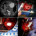
This is the case of a 78 years old patient with a history of acute coronary syndrome complicated by rupture of the left ventricle. The ventricular wall defect was closed by tachosyl heamostatic patchs and gluing. Two months after the occurrence of the acute coronary syndrom, computed tomography detected a huge apical pseudoaneurysm of the left ventricle ruptured into the pericardium (A). The patient underwent median sternotomy and cardiopulmonary bypass associated with a moderate hypothermia at 28°C, then, we performed the DOR technique for the cure of the pseudoaneurysm (B), using a Gore Tex patch and Teflon felt support (C,D).
Figure 1: A) computed tomography showing a huge apical pseudoaneurysm of the left ventricle with extravasation of the radiocontrast agent in the pericardium; B) a perioperative view showing the apical pseudoaneurysm of the left ventricle; C) a perioperative view showing the closure of the defect by a Gore Tex patch; D) a perioperative view showing the closure of the remaining re-entries by Teflon felt support
Search
This article authors
On Pubmed
On Google Scholar
Citation [Download]
Navigate this article
Similar articles in
Key words
Article metrics
Recently from the PAMJ


Authors´ services
