
Pycnodysostosis presented with tibial shaft fracture
Abdelhalim Mahmoudi, Youssef Bouabdallah
Corresponding author: Abdelhalim Mahmoudi, Department of Pediatric Surgery, University Hospital Hassan II, Fez, Morocco 
Received: 06 Dec 2015 - Accepted: 03 Jan 2016 - Published: 09 Jan 2020
Domain: Maternal and child health
Keywords: Pycnodysostosis, short stature, tibial shaft fracture
©Abdelhalim Mahmoudi et al. Pan African Medical Journal (ISSN: 1937-8688). This is an Open Access article distributed under the terms of the Creative Commons Attribution International 4.0 License (https://creativecommons.org/licenses/by/4.0/), which permits unrestricted use, distribution, and reproduction in any medium, provided the original work is properly cited.
Cite this article: Abdelhalim Mahmoudi et al. Pycnodysostosis presented with tibial shaft fracture. Pan African Medical Journal. 2020;35:5. [doi: 10.11604/pamj.2020.35.5.8581]
Available online at: https://www.panafrican-med-journal.com//content/article/35/5/full
Images in medicine 
Pycnodysostosis presented with tibial shaft fracture
Pycnodysostosis presented with tibial shaft fracture
Abdelhalim Mahmoudi1,&, Youssef Bouabdallah1
1Department of Pediatric Surgery, University Hospital Hassan II, Fez, Morocco
&Corresponding author
Abdelhalim Mahmoudi, Department of Pediatric Surgery, University Hospital Hassan II, Fez, Morocco

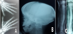
Pycnodysostosis is rare autosomal recessive lysosomal storage disorder of bone in which the enzyme Cathepsin K is mutated which maps on chromosome 1q21 causing osteoclast dysfunction. It is characterized by short stature, craniofacial dysmorphias, osteosclerosis, and brittle bones. Recognition of these signs is important in order to make the diagnosis and prevent possible complications. A 13-year old male patient of 130 cm height presented to the emergency with pathological fracture in shaft of left tibia. On the basis of clinical and radiological findings, he was diagnosed as a case of pycnodysostosis. The patient had history of pathological fracture in shaft of right tibia. With normal bone healing, open fontanelles, short stature, short finger tips, flat and grooved nails, micrognathia, and irregular teeth with hypodontia. Radiological evaluation of patient revealed generalized osteopetrosis, short distal phalanges, obtuse angle of mandible and open fontanelles. He was managed conservatively and showed normal bone healing pattern. No specific treatment options exist, so treatment is supportive, with fracture prevention and management constituting the most important aspects of clinical care. Dental hygiene and regular checkups are also helpful in preventing complications.
Figure 1: A) AP X-ray of both hands demonstrate short stubby fingers with dysplastic nails and acro-osteolysis; B) lateral skull X-rays reveal a large cranial vault with persistent open sutures and fontanelles as well as wormian bones: hypoplastic facial bones and an underdeveloped mandible with an obtuse jaw angle are also demonstrated; C) AP and lateral X-ray reveal a tibial shaft fracture
Search
This article authors
On Pubmed
On Google Scholar
Citation [Download]
Navigate this article
Similar articles in
Key words
Article metrics
PlumX Metrics
Pycnodysostosis presented with tibial shaft fractureRecently from the PAMJ


Authors´ services
