
Characterisation of Vibrio cholerae isolates from the 2009, 2010 and 2016 cholera outbreaks in Lusaka province, Zambia
Kapambwe Mwape, Geoffrey Kwenda, Annie Kalonda, John Mwaba, Chileshe Lukwesa-Musyani, Joseph Ngulube, Anthony Marius Smith, James Mwansa
Corresponding author: Kapambwe Mwape, Department of Basic Sciences, Michael Chilufya Sata School of Medicine, Copperbelt University, Ndola, Zambia 
Received: 30 Apr 2019 - Accepted: 29 Nov 2019 - Published: 07 Feb 2020
Domain: Bacteriology,Microbiology
Keywords: Cholera, Vibrio cholerae, multidrug resistance, Lusaka province
©Kapambwe Mwape et al. Pan African Medical Journal (ISSN: 1937-8688). This is an Open Access article distributed under the terms of the Creative Commons Attribution International 4.0 License (https://creativecommons.org/licenses/by/4.0/), which permits unrestricted use, distribution, and reproduction in any medium, provided the original work is properly cited.
Cite this article: Kapambwe Mwape et al. Characterisation of Vibrio cholerae isolates from the 2009, 2010 and 2016 cholera outbreaks in Lusaka province, Zambia. Pan African Medical Journal. 2020;35:32. [doi: 10.11604/pamj.2020.35.32.18853]
Available online at: https://www.panafrican-med-journal.com//content/article/35/32/full
Research 

Characterisation of Vibrio cholerae isolates from the 2009, 2010 and 2016 cholera outbreaks in Lusaka province, Zambia
Characterisation of Vibrio cholerae isolates from the 2009, 2010 and 2016 cholera outbreaks in Lusaka province, Zambia
Kapambwe Mwape1,2,&, Geoffrey Kwenda3, Annie Kalonda3, John Mwaba3,4, Chileshe Lukwesa-Musyani4, Joseph Ngulube4, Anthony Marius Smith5,6, James Mwansa7
1Department of Basic Sciences, Michael Chilufya Sata School of Medicine, Copperbelt University, Ndola, Zambia, 2Department of Pathology and Microbiology, School of Medicine, University of Zambia, Lusaka, Zambia, 3Department of Biomedical Sciences, School of Health Sciences, University of Zambia, Lusaka, Zambia, 4Department of Pathology and Microbiology, University Teaching Hospital, Lusaka, Zambia, 5Bacteriology Division, Centre for Enteric Diseases, National Institute for Communicable Diseases, National Health Laboratory Service, Johannesburg, South Africa, 6Department of Clinical Microbiology and Infectious Diseases, School of Pathology, Faculty of Health Sciences, University of the Witwatersrand, Johannesburg, South Africa, 7Department of Medical Microbiology, Faculty of Medicine, Lusaka Apex Medical University, Lusaka, Zambia
&Corresponding author
Kapambwe Mwape, Department of Basic Sciences, Michael Chilufya Sata School of Medicine, Copperbelt University, Ndola, Zambia
Introduction: in 2009 and 2010, more than 6,000 cholera cases were recorded during these outbreaks with more than 80% of cases recorded in Lusaka province. After a five-year break, in 2016 an outbreak occurred in Lusaka, causing more than 1,000 cases of cholera. This study will strengthen the epidemiological information on the changing characteristics of the cholera outbreaks, for treatment, prevention and control of the disease.
Methods: this was a laboratory-based descriptive cross-sectional study conducted at the University Teaching Hospital in Lusaka, Zambia. A total of 83 V. cholerae O1 isolates were characterised by biochemical testing, serotyping, antimicrobial susceptibility testing, and macrorestriction analysis using Pulsed-Field Gel Electrophoresis.
Results: macrorestriction analysis of the isolates demonstrated high genetic diversity among the isolates with 16 different patterns. The largest pattern comprised 9 isolates while the smallest one had 1 isolate. 2009 and 2010 isolates were highly resistant to nalidixic acid and cotrimoxazole, but highly sensitive to azithromycin and ampicillin. Of the fifty-two isolates from the 2016 cholera outbreak, 90% (47) were sensitive to cotrimoxazole, 94% (49) to tetracycline, and 98% (51) to azithromycin, while 98% (51) were resistant to nalidixic acid and 31(60%) to ampicillin.
Conclusion: macrorestriction analysis demonstrated high genetic diversity among the V. cholerae O1 strains, suggesting that these isolates were probably not from a similar source. This study also revealed the emergence of multidrug resistance among the 2016 V. cholerae outbreak isolates but were susceptible to cotrimoxazole, tetracycline, and azithromycin, which can be used for treatment of the cholera cases.

Cholera is an enteric disease of immense public health concern, causing an estimated 2.8 million cases with 91,000 deaths globally every year [1]. It is an acute disease characterised by severe watery diarrhoea caused by toxigenic Vibrio cholerae strains belonging to serogroups O1 and O139 [2]. The bacteria colonise the small intestine and produce an enterotoxin known as cholera toxin (CT) [3]. Pandemics that are caused by this bacterium have severely affected many countries on multiple continents for many years [4]. Cholera is transmitted via the faeco-oral route and is particularly associated with poverty and poor sanitation [3]. The first cholera outbreak in Africa was reported in 1836 along the Indian Ocean coast killing about 20,000 people [5]. No further outbreak was reported on the continent after the 1893-1894 outbreak in the Senegambia region until the seventh pandemic reached the continent in 1970 [6]. This pandemic caused massive outbreaks in Africa resulting in more than 400,000 cases with a high mortality rate [7]. Between 1995 and 2005, Africa experienced a greater upsurge in cholera outbreaks than other continents, with over 80% of the global total number of cholera cases [8]. The trend of Africa reporting more cholera cases continued between 2006 and 2010 [9]. In addition over the past 10 years, several Southern African countries, such as Mozambique [10], Tanzania [11], Zimbabwe [12], South Africa [2]and Zambia [13] have reported cholera outbreaks. Zambia usually experiences cholera outbreaks during the rainy season and most of them have been associated with fishing camps, especially in the northern part of the country and in unplanned settlements of Lusaka and Copperbelt Provinces [14]. The first cholera outbreak in Zambia was reported in the 1970s and several other outbreaks have occurred over the years, with the worst outbreak being in 1991 that resulted in over 13,000 cases [15]. In the 2009 outbreak, a total of 4,712 cases were reported, while the 2010 cholera outbreak caused 6,794 cases, with the majority of the cases being from Lusaka Province [13, 14]. In 2016 between February and June, Zambia experienced an outbreak with more than 1000 cases and 22 deaths being reported from Lusaka Province alone after a quiescent period of 5 years [16]. In order to minimize the disease burden caused by cholera, antimicrobial drugs have been used against V. cholerae O1 to shorten the duration of illness and to reduce the volume of stools produced [17]. However, the increased use of antibiotics against cholera has resulted in the emergence of multiple drug resistance in Zambia and other parts of the world [18-21]. During the 2009, 2010 and 2016 cholera outbreaks, Lusaka reported the highest number of cholera cases in Zambia [14]. Therefore, this study sought to describe the phenotypic characteristics of V. cholerae O1 strains isolated from Lusaka during these three cholera outbreaks through serotyping and antibiotic susceptibility testing, and their genetic diversity by macrorestriction analysis using Pulsed-Field Gel Electrophoresis (PFGE).

Identification and serotyping of the V. cholerae isolates: isolates used in this study were recovered from rectal swabs and stool samples from suspected cholera cases during the 2009, 2010 and 2016 cholera outbreaks in Lusaka district. After a positive cholera diagnosis by culture, the isolates of V. cholerae were stored in skimmed milk tryptone glucose glycerol (STGG) vials at -80�C. The isolates were revived by thawing the vial contents at room temperature and inoculating them in alkaline peptone water at 37�C for 6 hours. They were then subcultured onto Thiosulfate Citrate Bile Salts Sucrose (TCBS) agar and incubated at 37�C for 24 hours. The identity of the isolates was confirmed by biochemical tests as described in the Centers for Infectious Disease Control manual for cholera diagnosis (CDC, 2015) and serology using polyvalent O1 antisera and monovalent Inaba and Ogawa (Mast Diagnostics, Merseyside, United Kingdom) according to manufacturer´s instructions.
Macrorestriction analysis of the V. cholerae isolates: in order to determine the clonal relationships among the V. cholerae isolates, an analysis of chromosomal DNA restriction patterns was performed by Pulsed-Field Gel Electrophoresis (PFGE) with NotI digestion on a Bio-Rad CHEF-DR III electrophoretic system (Bio-Rad Laboratories, Hercules, USA) using a PulseNet protocol. PFGE patterns were analyzed using BioNumerics Version 6.5 Software (Applied Maths, Sint-Martens-Latem, Belgium) with dendrograms of the patterns created using the Unweighted Pair Group Method with Arithmetic Averages, with analysis of banding patterns incorporating the Dice-coefficient at an optimization setting of 1.5% and a position tolerance setting of 1.5%. Two or more isolates with a PFGE pattern percentage similarity value (index) of ≤ 92% were defined as clusters. Strains with pattern similarity value < 92% similarity were each assigned a distinct cluster number. Strains above 95% similarity value were considered identical.
Antibiotic susceptibility testing of the V. cholerae isolates: antibiotic susceptibility was determined using the Bauer-Kirby disk diffusion test. All strains were tested for resistance to ampicillin (10μg), ciprofloxacin (5μg), tetracycline (30μg), trimethoprim-sulphamethoxazole (25μg) and Nalidixic acid (30μg) using commercially available discs (Oxoid Limited, Hampshire, United Kingdom). Minimum inhibitory concentrations (MIC) of azithromycin was determined using E-test strips (bioMérieux, Marcy I´Etoile, France) according to manufacturer's instructions. Breakpoints for assessing resistance were determined following the Clinical Laboratory Standards Institute (CLSI) guidelines M45 document [22].
Ethics approval: this study was laboratory-based and involved no direct contact with patients. All participant specimens were de-identified and given study-specific identification codes. Permission to conduct the study was sought from the University Teaching Hospital Management in Lusaka, whilst ethics clearance was sought from the University of Zambia Biomedical Research and Ethics Committee (UNZABREC). The ethics clearance certificate number was 014-11-15.
Identification and serotyping of the V. cholerae isolates: a total of 83 isolates were successfully revived and were all identified as V. cholerae O1. Out of these, 6 isolates were from the 2009 outbreak, while 25 were from the 2010 and 52 from the 2016 outbreaks. The Ogawa serotype accounted for 70% (58), which were from the 2009 and 2016 outbreaks, while the Inaba serotype constituted 30% (25), all of which were from the 2010 outbreak, of the isolates.
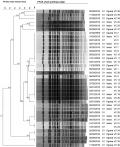
Macrorestriction analysis of the V. cholerae isolates: PFGE analysis demonstrated high diversity amongst the 38 isolates analysed. When electrophoretic profiles were analysed, 6 clusters, with at least 92% similarity, could be observed (Figure 1). This similarity index (SI) was determined by comparing the three possible cut-offs, 90%, 92%, and 95%. The SI cut-off that provided the best discrimination, after a visual inspection was 92% as it correctly assigned the isolates to their respective clusters. Six clusters were produced with the largest cluster containing (12/38) strains which were 97.8 to 100% similar, followed by a cluster of 8 strains (98 to 100% similarity), then a cluster of 7 strains (98.1 to 100% similarity) and a cluster 4 strains (98 to 100% similarity). The smallest clusters contained 3 strains (93.8 to 95.8% similarity) and (95.3 to 98% similarity). Only one isolate, VC40, remained outside the clusters.
Antibiotic susceptibility testing of the V. cholerae isolates: all the 2009 and 2010 isolates (31/83) were 100% resistant to nalidixic acid and cotrimoxazole but were all sensitive to ampicillin (100%) and azithromycin (100%). Reduced sensitivity to tetracycline for both the 2009 (67%) and 2010 (64%) isolates was observed, with most of them (65%) falling in the intermediate category. Out of the 52 isolates from the 2016 outbreak, 98% (51/52) were resistant to nalidixic acid. The majority of these isolates were highly sensitive to cotrimoxazole (90%, 47/52), tetracycline (98%, 51/52) and azithromycin (98%, 51/52). Interestingly, reduced sensitivity to ciprofloxacin (6%, 3/52) was observed with most of the isolates (83%, 43/52) falling in the intermediate category for this antibiotic. An attempt was also made to profile antimicrobial resistance (AMR) patterns of the bacterial isolates. All of the outbreaks exhibited AMR but very low multidrug resistance (MDR) was detected. None of the 2009 (0%, 0/6) and the 2010 (0%, 0/25) outbreak isolates were MDR while one of the 2016 outbreak isolates (1.9%, 1/52) displayed an MDR pattern of Tetracycline-Trimethoprim-Sulfamethoxazole-Ampicillin-Azithromycin (Table 1). MDR was defined as resistance to three or more drugs from different classes [23].
Cholera outbreaks continue to be major public health threat in Lusaka Province. These outbreaks usually occur in peri-urban townships that are densely populated and have inadequate water and sanitation facilities, a scenario that favours the spread of the disease. The sources of infection have been attributed to contaminated water supplies, contaminated food, inadequate sanitation and poor hygienic practices. Most of these outbreaks occur in the rainy season, thereby predisposing people to the risk of contracting the disease. Data presented in this study demonstrate that V. cholerae O1 strains belonging to the Ogawa serotype were responsible for both the 2009 and 2016 outbreaks, while the Inaba serotype was responsible for the 2010 outbreak. Several previous studies have demonstrated that the serotypes Ogawa and Inaba are the most encountered serotypes in other African countries, such as South Africa [2], Tanzania [11], Mozambique [24], Kenya [25] and Ghana [26]. However, a study in the Democratic Republic of Congo (DRC) reported some strains belonging to another serotype, Hikojima, during a 2009 cholera outbreak there [27]. It is interesting to note that a different serotype was reported from the DRC, a country that shares a long border area with Zambia on its southern side. This calls for vigilance against the Hikojima serotype, especially during screening exercises for cholera cases that may arise from this border area. Molecular epidemiology of human bacterial pathogens provides valuable information for understanding the reservoir, pathogenicity and control of bacterial pathogens [28].
Macrorestriction analysis data revealed 16 patterns among all the isolates, with those from 2009 and 2010 outbreak clustering together. This suggests that these isolates were closely related and that those from the 2009 outbreak may have continued to cause infection during the 2010 cholera outbreak. This phenomenon has also been reported in Kenya [25], South Africa [29] and India [30]. The 2016 isolates, however, formed unique clusters, suggesting the emergence of a new clade of the Ogawa serotype causing the cholera outbreak in Lusaka. Similar findings were also reported in a previous Zambian study on outbreak strains isolated between 1996 and 2004 in Zambia, which revealed the emergence of new V. cholerae strains [31]. Yet another study in Haiti showed the same phenomenon [32]. These findings suggest that the evolution of V. cholerae led to a shift between serotypes Inaba and Ogawa [33]. The strains in this current study generally exhibited high genetic diversity with one isolate, from the 2016 outbreak, not showing any relationship with any of the other isolates. This high genetic diversity seems to suggest that the isolates did not have the same origin [34]. The increase in AMR among enteropathogens, especially MDR strains, is challenging global prospects for fighting diarrhoeal disease pathogens. The emergence and spread of MDR pathogens outstrips the development of drugs, shrinking the therapeutic arsenal [35]. In this study, all the organisms were totally resistant to nalidixic acid and partially resistant to ampicillin. A previous Zambian study, during the 1990-1991 major cholera outbreak, showed low resistance of V. cholerae O1 strains to cotrimoxazole, tetracycline, chloramphenicol and doxycycline but significant increase in AMR during subsequent outbreaks [18].
The high resistance observed with cotrimoxazole supports our findings among the 2009 and 2010 isolates, but interestingly very low resistance to this drug was observed among the 2016 isolates. In consonance with our findings from all the three outbreaks, a Nepalese study reported 100% resistance to nalidixic acid [36]. High rates of sensitivity to azithromycin were also reported in a study carried out in Iran, which was in accordance with our findings where only 1.2% resistance was reported [37]. Unlike the findings in this study, high resistance levels to tetracycline (82%) were reported in Mozambique [24]. In this current study, the emergence of MDR strains was observed with the 2016 outbreak where one of the isolates was MDR. These findings were in contrast with the findings of a study conducted in Cameroon where the MDR detection rate was 92% [38]. Strains with new resistance patterns also emerged with reduced sensitivity to ciprofloxacin among the 2016 isolates. V. cholera O1 strains with reduced quinolone sensitivity in Africa were first reported in a Zimbabwean study [39] and later similar findings were reported in Nigeria [40], Kenya [25] and Ghana [26]. Differences in the antibiotic resistance patterns observed during the three outbreaks and the emergence of MDR strains may be attributed to a spontaneous mutation that results from the abuse of antibiotics and horizontal gene transfer [41].
Data in this study demonstrate that the 2009, 2010 and 2016 cholera outbreaks were caused by V. cholerae serogroup O1, with the Ogawa serotype being the predominant serotype. Serotype switching may have occurred in the three outbreaks. However, it is unclear as to what mechanisms could have triggered this process. Another significant finding was that V. cholerae strains showed high genetic diversity and therefore, could not have come from a single source of infection. This calls for tracking of the sources of infection as an effective means of controlling cholera in affected areas of Lusaka or other parts of the country. The strains that caused the 2009 cholera outbreak also emerged during the 2010 outbreak but new strains of the bacteria emerged during the 2016 outbreak as evidenced by the macrorestriction and AMR patterns. This study also revealed the emergence of MDR isolates, which were obtained from the 2016 outbreak, with ampicillin and nalidixic acid being the most ineffective drugs. Cotrimoxazole and nalidixic acid were the most ineffective drugs for the 2009 and 2010 isolates. An interesting observation was the reversion to cotrimoxazole sensitivity and reduced quinolone sensitivity for the 2016 isolates. Despite the detection of AMR in all three outbreaks and MDR in the 2016 isolates, cheaper antibiotics such as cotrimoxazole, tetracycline and azithromycin proved to be potent drugs for cholera treatment. This study presents data that underscores the need for close monitoring of V. cholerae strains that cause cholera outbreaks in Zambia. This is important to ensure that the correct antibiotic is chosen according to resistance variations, considering the increasing global burden of cholera, and the emergence and spread of new variants that will significantly influence the clinical management of cholera and its prevention.
What is known about this topic
- Cholera is caused by Vibrio cholerae serogroups O1/O139 and serotypes Inaba, Ogawa and Hikojima;
- Emergence of multi-drug resistance has been reported elsewhere;
- Cholera outbreaks in an area can either be caused by highly related or diverse strains.
What this study adds
- Cholera outbreaks in Lusaka province were due to Vibrio cholerae O1 serotype Ogawa in 2009, Inaba in 2010 and Ogawa in 2016;
- Antibiotic resistant strains were circulating in all three outbreaks in Lusaka province but the 2016 outbreak demonstrated emergence of multidrug resistance;
- Macrorestriction analysis demonstrated emergence of new Vibrio cholerae variants during the 2016 outbreak in Lusaka province as they formed unique clusters.
The authors declare no competing interests.
KM, JM and GK, contributed to the conceptualisation, investigation, data analysis and writing of the manuscript. AK, CLM and JN contributed to the investigation and writing of the manuscript. JM contributed to the conceptualisation, and writing of the manuscript. AMS, contributed to the investigation, data analysis and writing of the manuscript.
The authors are grateful to the University Teaching Hospital (UTH) management for allowing them to conduct the study using the UTH facilities. We would also like to acknowledge the Copperbelt University for the financial assistance for KM during the study. We are also grateful to the National Institute for Communicable Diseases in Johannesburg, South Africa for facilitating the molecular analysis of the V. cholerae isolates.
Table 1: antimicrobial resistance profile of V. cholerae O1 isolates from the 2009, 2010 and 2016 cholera outbreaks
Figure 1: genetic relationships amongst the PFGE NotI macrorestriction profiles of V. cholerae O1 isolates from Lusaka Province
- Ali M, Nelson AR, Lopez AL, Sack DA. Updated global burden of cholera in endemic countries. PLoS Negl Trop Dis. 2015 Jun 4;9(6):e0003832. eCollection 2015. PubMed | Google Scholar
- Ismail H, Smith AM, Tau NP, Sooka A, Keddy KH, Group for Enteric R. Meningeal Disease Surveillance in South Africa: Cholera outbreak in South Africa, 2008-2009: laboratory analysis of Vibrio cholerae O1 strains. J Infect Dis. 2013 Nov 1;208 Suppl 1:S39-45. PubMed | Google Scholar
- Oguttu DW, Okullo A, Bwire G, Nsubuga P, Ario AR. Cholera outbreak caused by drinking lake water contaminated with human faeces in Kaiso Village, Hoima District, Western Uganda, October 2015. Infect Dis Poverty. 2017 Oct 10;6(1):146. PubMed | Google Scholar
- Ramazanzadeh R, Rouhi S, Shakib P, Shahbazi B, Bidarpour F, Karimi M. Molecular Characterization of Vibrio cholerae Isolated From Clinical Samples in Kurdistan Province, Iran. Jundishapur J Microbiol. 2015 May 31;8(5):e18119. eCollection 2015 May. PubMed | Google Scholar
- Olago D, Marshall M, Wandiga SO, Opondo M, Yanda PZ, Kanalawe R et al. Climatic, socio-economic, and health factors affecting human vulnerability to cholera in the Lake Victoria basin, East Africa. Ambio. 2007 Jun;36(4):350-8. PubMed | Google Scholar
- Mengel MA, Delrieu I, Heyerdahl L, Gessner BD. Cholera outbreaks in Africa. Current topics in microbiology and immunology. 2014;379:117-144. PubMed | Google Scholar
- Harris JB, LaRocque RC, Qadri F, Ryan ET, Calderwood SB. Cholera. Lancet. 2012;379(9835):2466-2476. PubMed
- Griffith DC, Kelly-Hope LA, Miller MA. Review of reported cholera outbreaks worldwide, 1995-2005. The American journal of tropical medicine and hygiene. 2006;75(5):973-977. PubMed | Google Scholar
- Saidi SM, Chowdhury N, Awasthi SP, Asakura M, Hinenoya A, Iijima Y et al. Prevalence of Vibrio cholerae O1 El Tor variant in a cholera-endemic zone of Kenya. J Med Microbiol. 2014 Mar;63(Pt 3):415-20. PubMed| Google Scholar
- Gujral L, Sema C, Rebaudet S, Taibo CL, Manjate AA, Piarroux R et al. Cholera epidemiology in Mozambique using national surveillance data. J Infect Dis. 2013 Nov 1;208 Suppl 1:S107-14. PubMed | Google Scholar
- Kachwamba Y, Mohammed AA, Lukupulo H, Urio L, Majigo M, Mosha F et al. Genetic Characterization of Vibrio cholerae O1 isolates from outbreaks between 2011 and 2015 in Tanzania. BMC infectious diseases. 2017;17(1):157. PubMed | Google Scholar
- Maponga BA, Chirundu D, Gombe NT, Tshimanga M, Bangure D, Takundwa. L. Cholera: a comparison of the 2008-9 and 2010 outbreaks in Kadoma City, Zimbabwe. The Pan African medical journal. 2015;20:221. PubMed | Google Scholar
- Olu O, Babaniyi O, Songolo P, Matapo B, Chizema E, Kapin'a-Kanyanga M et al. Cholera Epidemiology in Zambia from 2000 to 2010: Implications for Improving Cholera Prevention and Control Strategies in the Country. East African medical journal. 2013;90(10):324-331. PubMed | Google Scholar
- Zambia Ministry of Health: Cholera Control Guidelines- First Edition. 2011. Lusaka, Zambia.
- DuBois AE, Sinkala M, Kalluri P, Makasa-Chikoya M, Quick RE. Epidemic cholera in urban Zambia: hand soap and dried fish as protective factors. Epidemiology and infection. 2006;134(6):1226-1230. PubMed | Google Scholar
- Sladoje M. International Growth Centre Zambia: an institutional perspective. 2018. Accessed 10 April 2019.
- Kitaoka M, Miyata ST, Unterweger D, Pukatzki S. Antibiotic resistance mechanisms of Vibrio cholerae. Journal of medical microbiology. 2011;60(4):397-407. PubMed | Google Scholar
- Mwansa JC, Mwaba J, Lukwesa C, Bhuiyan NA, Ansaruzzaman M, Ramamurthy T et al. Multiply antibiotic-resistant Vibrio cholerae O1 biotype El Tor strains emerge during cholera outbreaks in Zambia. Epidemiology and infection. 2007;135(5):847-853. PubMed | Google Scholar
- Rashed SM, Mannan SB, Johura FT, Islam MT, Sadique A, Watanabe H et al. Genetic characteristics of drug-resistant Vibrio cholerae O1 causing endemic cholera in Dhaka, 2006-2011. Journal of medical microbiology. 2012;61(12):1736-1745. PubMed | Google Scholar
- Mahmud ZH, Islam S, Zaman RU, Akter M, Talukder KA, Bardhan PK et al. Phenotypic and genotypic characteristics of Vibrio cholerae O1 isolated from the Sierra Leone cholera outbreak in 2012. Transactions of the Royal Society of Tropical Medicine and Hygiene. 2014;108(11):715-20. PubMed | Google Scholar
- Jain M, Kumar P, Goel AK. Emergence of Tetracycline Resistant Vibrio cholerae O1 Biotype El Tor Serotype Ogawa with Classical ctxB Gene from a Cholera Outbreak in Odisha, Eastern India. Journal of pathogens. 2016;2016:1695410. PubMed | Google Scholar
- CLSI. Methods for Antimicrobial Dilution and Disk Susceptibility Testing of Infrequently Isolated or Fastidious Bacteria- 3rd edition. 2016. CLSI.
- Magiorakos AP, Srinivasan A, Carey RB, Carmeli Y, Falagas ME, Giske CG et al. Multidrug-resistant, extensively drug-resistant and pandrug-resistant bacteria: an international expert proposal for interim standard definitions for acquired resistance. Clinical microbiology and infection : the official publication of the European Society of Clinical Microbiology and Infectious Diseases 2012.18(3):268-281. PubMed | Google Scholar
- Dengo-Baloi LC, Sema-Baltazar CA, Manhique LV, Chitio JE, Inguane DL, Langa JP. Antibiotics resistance in El Tor Vibrio cholerae 01 isolated during cholera outbreaks in Mozambique from 2012 to 2015. PloS one. 2017;12(8):e0181496. PubMed | Google Scholar
- Mercy N, Mohamed AA, Zipporah N, Chowdhury G, Pazhani GP, Ramamurthy T et al. Phenotypic and genetic characterization of Vibrio cholerae O1 isolated from various regions of Kenya between 2007 and 2010. The Pan African medical journal.2014;19:8. PubMed | Google Scholar
- Eibach D, Herrera-Leon S, Gil H, Hogan B, Ehlkes L, Adjabeng M et al. Molecular Epidemiology and Antibiotic Susceptibility of Vibrio cholerae Associated with a Large Cholera Outbreak in Ghana in 2014. PLoS neglected tropical diseases. 2016;10(5):e0004751. PubMed | Google Scholar
- Miwanda B, Moore S, Muyembe JJ, Nguefack-Tsague G, Kabangwa IK, Ndjakani DY et al. Antimicrobial Drug Resistance of Vibrio cholerae, Democratic Republic of the Congo. Emerging infectious diseases. 2015;21(5):847-851. PubMed | Google Scholar
- De R, Ghosh JB, Sen Gupta S, Takeda Y, Nair GB. The role of Vibrio cholerae genotyping in Africa. The Journal of infectious diseases. 2013;208(1):S32-38. PubMed | Google Scholar
- Keddy KH, Nadan S, Govind C, Sturm AW. Group for Enteric R, Meningeal Disease Surveillance in South A: Evidence for a clonally different origin of the two cholera epidemics of 2001-2002 and 1980-1987 in South Africa. Journal of medical microbiology. 2007;56(12):1644-1650. Google Scholar
- Torane V, Kuyare S, Nataraj G, Mehta P, Dutta S, Sarkar B. Phenotypic and antibiogram pattern of V. cholerae isolates from a tertiary care hospital in Mumbai during 2004-2013: a retrospective cross-sectional study. BMJ open. 2016;6(11):e012638. PubMed | Google Scholar
- Bhuiyan NA, Nusrin S, Ansaruzzaman M, Islam A, Sultana M, Alam M et al. Genetic characterization of Vibrio cholerae O1 strains isolated in Zambia during 1996-2004 possessing the unique VSP-II region of El Tor variant. Epidemiology and infection. 2012,140(3):510-518. PubMed | Google Scholar
- Alam MT, Ray SS, Chun CN, Chowdhury ZG, Rashid MH, Madsen Beau De Rochars VE et al. cholerae O1 from Ogawa to Inaba Serotype Isolated from Clinical and Environmental Samples in Haiti. PLoS neglected tropical diseases. 2016;10(10):e0005045. PubMed | Google Scholar
- Lu H, Yuan Y, Sun N, Bi Z, Guan B, Shao K et al. Characterization of Vibrio cholerae isolates from 1976 to 2013 in Shandong Province, China. Brazilian journal of microbiology : [publication of the Brazilian Society for Microbiology]. 2017;48(1):173-179. PubMed | Google Scholar
- Hasan NA, Choi SY, Eppinger M, Clark PW, Chen A, Alam M et al. Genomic diversity of 2010 Haitian cholera outbreak strains. Proceedings of the National Academy of Sciences of the United States of America. 2012;109(29):E2010-2017. PubMed | Google Scholar
- Dantas G SM. How to Fight Back Against Antibiotic Resistance. American Scientist. 2014;102(1):42-51. Google Scholar
- Dixit SM, Johura FT, Manandhar S, Sadique A, Rajbhandari RM, Mannan SB et al. Cholera outbreaks (2012) in three districts of Nepal reveal clonal transmission of multi-drug resistant Vibrio cholerae O1. BMC infectious diseases. 2014;14:392. PubMed | Google Scholar
- Barati H, Moradi G, Rasouli MA, Mohammadi P. Epidemiologic and Drug Resistance Pattern of Vibrio cholerae O1 Biotype El Tor, Serotype Ogawa, in the 2011 Cholera Outbreak, in Alborz Province, Iran. Jundishapur journal of microbiology. 2015;8(11):e23477. PubMed | Google Scholar
- Akoachere Kihla JFT, Masalla TN, Njom HA. Multi-drug resistant toxigenic Vibrio cholerae O1 is persistent in water sources in New Bell-Douala, Cameroon. BMC infectious diseases. 2013:13;366. PubMed | Google Scholar
- Islam MS, Midzi SM, Charimari L, Cravioto A, Endtz HP. Susceptibility to fluoroquinolones of Vibrio cholerae O1 isolated from diarrheal patients in Zimbabwe. Jama. 2009;302(21):2321-2322. PubMed | Google Scholar
- Marin MA, Thompson CC, Freitas FS, Fonseca EL, Aboderin AO, Zailani SB et al. Cholera outbreaks in Nigeria are associated with multidrug resistant atypical El Tor and non-O1/non-O139 Vibrio cholerae. PLoS neglected tropical diseases. 2013;7(2):e2049. PubMed | Google Scholar
- Thapa Shrestha U, Adhikari N, Maharjan R, Banjara MR, Rijal KR, Basnyat SR et al. Multidrug resistant Vibrio cholerae O1 from clinical and environmental samples in Kathmandu city. BMC infectious diseases. 2015;15:104. PubMed | Google Scholar
Search
This article authors
On Pubmed
On Google Scholar
Citation [Download]
Navigate this article
Similar articles in
Key words
Tables and figures

Article metrics
Recently from the PAMJ


Authors´ services
