
A post traumatic capsulorhexis
Kawtar Belkhadir, Ouafaa Cherkaoui
Corresponding author: Kawtar Belkhadir, Departement d´Ophtalmologie Unit A, Hopital des Spécialités, Faculté de Médicine et Pharmacie, Université Mohammed V, Rabat, Maroc 
Received: 18 Oct 2019 - Accepted: 01 Nov 2019 - Published: 20 Apr 2020
Domain: Urgent Care Medicine,Ophthalmology
Keywords: Ocular trauma, post traumatic cataract, capsulorhexis
©Kawtar Belkhadir et al. Pan African Medical Journal (ISSN: 1937-8688). This is an Open Access article distributed under the terms of the Creative Commons Attribution International 4.0 License (https://creativecommons.org/licenses/by/4.0/), which permits unrestricted use, distribution, and reproduction in any medium, provided the original work is properly cited.
Cite this article: Kawtar Belkhadir et al. A post traumatic capsulorhexis. Pan African Medical Journal. 2020;35:134. [doi: 10.11604/pamj.2020.35.134.20705]
Available online at: https://www.panafrican-med-journal.com//content/article/35/134/full
Images in medicine 
A post traumatic capsulorhexis
A post traumatic capsulorhexis
Kawtar Belkhadir1, Ouafaa Cherkaoui1
1D�partement d´Ophtalmologie Unit A, H�pital des Spécialités, Faculté de Médicine et Pharmacie, Université Mohammed V, Rabat, Maroc
&Corresponding author
Kawtar Belkhadir, D�partement d´Ophtalmologie Unit A, H�pital des Spécialités, Faculté de Médicine et Pharmacie, Université Mohammed V, Rabat, Maroc
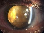
We report the case of a patient aged 50 years, with no pathological history, who consulted for a decrease in visual acuity secondary to an ocular trauma of the left eye occurred 6 months ago. The clinical examination found a visual acuity reduced to luminous perception, a clear cornea, an anterior chamber of good depth. Examination after pupillary dilation revealed an anterior capsule broken, in the manner of an incomplete circular anterior capsulorhexis, with retraction of the anterior capsule in superior temporal area, and yellowish pupillary reflection. The examination was completed by a B-mode ultrasonography which revealed a vitreous dislocated lens nucleus with vitreous organization in favor of intravitreal hemorrhage secondary to the trauma associated with an old retinal detachment. In view of the severity and the age of the post-traumatic lesions, therapeutic abstention was advocated. Ocular contusion trauma is a common cause of ophthalmic emergency consultation. An early and complete examination is necessary in order to carry out an exhaustive lesion assessment and to allow an early and adequate management in order to avoid the potentially blinding sequelae of these affections.
Figure 1: image of the left eye showing a retraction of anterior capsule
Search
This article authors
On Pubmed
On Google Scholar
Citation [Download]
Navigate this article
Similar articles in
Key words
Tables and figures
Article metrics
PlumX Metrics
A post traumatic capsulorhexisRecently from the PAMJ


Authors´ services
