
Puberté précoce chez une fillette révélant un corticosurrénalome malin
Ayad Anass, Amina Kili
Corresponding author: Ayad Anass, Service d’Hématologie et Oncologie Pédiatrique, Hôpital d’Enfants de Rabat, Maroc 
Received: 03 Apr 2017 - Accepted: 03 Apr 2017 - Published: 02 May 2017
Domain: Pediatric oncology
Keywords: Corticosurrénalome, puberté, chimiothérapie, masse
©Ayad Anass et al. Pan African Medical Journal (ISSN: 1937-8688). This is an Open Access article distributed under the terms of the Creative Commons Attribution International 4.0 License (https://creativecommons.org/licenses/by/4.0/), which permits unrestricted use, distribution, and reproduction in any medium, provided the original work is properly cited.
Cite this article: Ayad Anass et al. Puberté précoce chez une fillette révélant un corticosurrénalome malin. Pan African Medical Journal. 2017;27:6. [doi: 10.11604/pamj.2017.27.6.12412]
Available online at: https://www.panafrican-med-journal.com//content/article/27/6/full
Original article 

Puberté précoce chez une fillette révélant un corticosurrénalome malin
Puberté précoce chez une fillette révélant un corticosurrénalome malin
Precocious puberty in a little girl revealing malignant corticosurrenaloma
Ayad Anass1,&, Amina Kili2
1Service d’Hématologie et Oncologie Pédiatrique, Hôpital d’Enfants de Rabat, Maroc, 2Service d’Hématologie et Oncologie Pédiatrique, Hôpital d’Enfants de Rabat, Maroc
&Auteur correspondant
Ayad Anass, Service d’Hématologie et Oncologie Pédiatrique, Hôpital d’Enfants de Rabat, Maroc
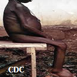
We report the case of a 5-year old little girl hospitalized for heterosexual precocious puberty. She was born by cesarean section and her first cry was immediate; she weighed 4 kg at birth, her psychomotor development was normal and she had no family history of malignancy. She had the onset of her disease at age 4. It was characterized by the occurrence of progressive pubic hair, acne, seborrhea, clitoral hypertrophy, a deeper voice as well as by the development of male muscle mass and the appearance of hair on her upper lip and face. These symptoms were associated with aggressive and agitated behavior. Abdominal palpation showed huge mass difficult to delineate, extending from the hypochondrium to the right flank reaching the umbilicus. Physical examination of the external genitalia showed large lips, absence of small lips, clitoral hypertrophy (penile clitoris) and stage 4 pubic hair (according to Tanner's classification). The remainder of the physical examination was normal. Hormonal assessment showed elevation of aldosterone, ΔAndrostendione, 17 hydroxyprogesterone, SDHA and testosterone as well as an 08.00 hours plasma cortisol level of 778,5 nmol/l (VN= 280-876nmol/l). Abdominal CT scan showed heterogeneous poly- lobed adrenal mass with hypervascular malignant-like necrotic areas, most likely corticosurrenaloma. The little girl underwent complete tumor resection and anatomopathologic confirmation. The postoperative course was uneventful. She underwent hydrocortisone therapy at a dose of 15 mg/m˛/i associated with mineralocorticoids. No chemotherapy was administered.
Key words: Corticosurrenaloma, puberty, chemotherapy, mass

Il s'agit d'une fillette de 5ans, hospitalisée pour puberté précoce hétérosexuelle. Elle est née par césarienne, le premier cri était immédiat avec un poids de 4 kg, le développement psychomoteur était normal et pas d'histoire de malignité dans la famille. Le début de sa maladie remontait ŕ l'âge de 4 ans par l'apparition d'une pilosité pubienne progressive, d'une acné, de séborrhée et d'une hypertrophie clitoridienne, une voix grave et développement d'une masse musculaire de type masculin. Ainsi que l'apparition de duvet au niveau de la lčvre supérieure et sur le visage. Le tout évoluant dans un contexte d'agitation et d'agressivité. La palpation abdominale, a trouvé une énorme masse difficile ŕ délimiter, allant de l'hypochondre jusqu'au flanc droit et arrivant jusqu'ŕ l'ombilic. L'examen des organes génitaux externes a noté la présence des grandes lčvres. Absence des petites lčvres et hypertrophie clitoridienne (clitoris pénien) et une pilosité pubienne stade 4 selon la classification de Tanner. Le reste de l'examen somatique était normal. Le bilan hormonal a objectivé une élévation de l'aldostérone, Δ Androstendione, 17 hydroxyprogéstérone, SDHA, et la testosterone ainsi qu'une cortisolémie de 8 h ŕ 778,5 nmol/l (VN= 280-876nmol/l). La TDM abdominale a montré une masse d'origine surrénalienne, hétérogčne, polylobé avec des zones de nécrose, hyper vasculaires, d'allure maligne corticosurrénalome trčs probable. Une exérčse tumorale totale a été réalisée avec confirmation anatomopathologique, Les suites opératoires étaient simples. La petite a été mise sous hydrocortisone ŕ la dose 15 mg/m2/j associés ŕ un minéralocorticoďde. Aucune chimiothérapie n'a été donnée.
Figure 1: image de la masse abdominale avec signes de puberté précoce
Search
This article authors
On Pubmed
On Google Scholar
Citation [Download]
Navigate this article
Similar articles in
Key words
Tables and figures

Article metrics
Recently from the PAMJ


Authors´ services
