
An unusual location of a cavernous hemangioma: a case report
Zahra Sayad, Bouchra Dani, Salma Benazzou, Malik Boulaadas
Corresponding author: Zahra Sayad, Department of Oral and Maxillofacial Surgery, Ibn Sina University Hospital Center, Rabat, Morocco 
Received: 21 Feb 2021 - Accepted: 23 Mar 2021 - Published: 11 May 2021
Domain: Maxillofacial surgery
Keywords: Cavernous, hemangioma, diagnostic, treatment, case report
©Zahra Sayad et al. Pan African Medical Journal (ISSN: 1937-8688). This is an Open Access article distributed under the terms of the Creative Commons Attribution International 4.0 License (https://creativecommons.org/licenses/by/4.0/), which permits unrestricted use, distribution, and reproduction in any medium, provided the original work is properly cited.
Cite this article: Zahra Sayad et al. An unusual location of a cavernous hemangioma: a case report. Pan African Medical Journal. 2021;39:29. [doi: 10.11604/pamj.2021.39.29.28492]
Available online at: https://www.panafrican-med-journal.com//content/article/39/29/full
Case report 

An unusual location of a cavernous hemangioma: a case report
An unusual location of a cavernous hemangioma: a case report
Zahra Sayad1,&, Bouchra Dani1, Salma Benazzou1, Malik Boulaadas1
&Corresponding author
Hemangiomas are benign vascular tumors that most often affect the skin, mucous membranes, subcutaneous tissues, bone and on rare occasions muscles. In the head and neck region, the masseter and trapezius muscles are most often affected; the temporalis muscle involvement is extremely rare. It is a childhood pathology that rarely occurs in adults. We report a case of a cavernous hemangioma in a 37-year-old female. Through this case and in the light of literature we focus on the clinicopathological aspects of this tumor and the rarity of this location.

Intramuscular hemangiomas are benign vascular neoplasms that represent less than 1% of all hemangiomas and which are often localized in the trunk and extremities. There are three types of hemangioma depending on the size of the affected vessel: capillary hemangioma, cavernous hemangioma, and compound hemangioma [1]. We report a case of a rare location of a cavernous hemangioma in the temporalis muscle with an extension to the infra temporal fossa.

A 37-year-old female, without significant personal or family medical history. The patient presents a swelling of the left temporal fossa that has been evolving for 5 years by gradually increasing in volume leading to a facial asymmetry. The physical examination revealed a soft, painless, non-pulsatile mass (4 x 2.5 cm in size), without thrill, not increasing volume in the declivity position, fixed to the deep plane, mobile to the skin, without inflammatory signs or lymphadenopathy.
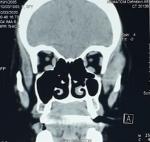
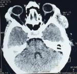
Contrast-enhanced computed tomography was performed, showing a temporalis muscle mass that extends to the infra-temporal fossa, measuring 33x17x39mm, isodense, unencapsulated, with polylobed contours, that enhanced after contrast injection and respected the subcutaneous fat (Figure 1, Figure 2).
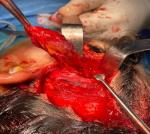
The patient underwent surgery, where we performed a hemi-coronal incision. The mass was found in the temporalis muscle and was excised completely without damaging the frontalis branch of the facial nerve. The operating follow-ups were simple, with excellent functional and aesthetic results (Figure 3).
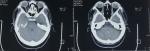
Macroscopic mass examination showed a smooth, nodular tumor tissue measuring 5 x 2.5 x 1 cm, with a purplish-brownish appearance. The histological study found a benign proliferation of dilated blood vessels with variable shape and size often cavernous that were trapped between muscle fibers; they are bordered by a well differentiated endothelium and supported by fibrous interstitial tissue in favor of cavernous hemangioma. Over a two-year period, no signs of recurrence were detected (Figure 4).
Hemangiomas are benign tumors characterized by abnormal proliferation of blood vessels. Their intramuscular locations represent less than 1% of all hemangiomas. Only 14% are located at the head and neck region. Masseter (36%), trapezius (24%) and sternocleidomastoid muscles are the most affected, while temporal muscle involvement remains extremely rare. To our knowledge, only 29 cases have been reported in the literature. A slight female predominance was described with an average age of 33 years [1, 2].
The etiopathogenesis of these tumors are unknown, yet there are several theories: congenital, hormonal factors and trauma. There are three types depending on the size of the vessel involved: capillary (small vessels) most common with an estimated incidence of 68%, Cavernous (26%) (Large vessels) followed by compound types (6%). Cavernous hemangioma is most frequently found in the temporal muscle [2, 3]. Clinically, they appear as a slow growing mass, mobile, painless, without pulsation, and without staining of the skin. The differential diagnosis includes lipoma, neurofibroma, dermoid cyst, enlarged lymph nodes, soft-tissue sarcoma, myositis ossificans and temporal arteritis [1, 4].
Imaging help to make a positive diagnosis and guiding the management of this tumor. Computed tomography is useful for defining the form, size, eventual bone damage, and infiltration of the surrounding tissues. However, MRI is the method of choice in defining the vascular nature of the tumor and providing further information on the exact extent of the tumor. Hemangiomas are generally isointense to muscle on T1-weighted images and hyperintense on T2-weighted images. Arteriography allows to identify the feeding vessels of the mass for a preoperative embolization if necessary [1, 3, 5].
Several therapeutic modalities are available ranging from simple observation, injection of sclerosing agents, corticosteroid treatment, radiotherapy, embolization (especially in preoperative) to reduce intraoperative bleeding, arriving at complete surgery excision which remains the method of choice in the definitive treatment of this tumor. In some cases of voluminous tumor, injection of sclerosing agents, corticosteroid treatment and radiotherapy, can be indicated as alternatives or adjuvants to surgery [6, 7]. In our case, under general anesthesia, hemi-coronal incision is used to control the region and the careful surgical dissection allows to prevent injury of the temporal and auricular branches of the facial nerve. The therapeutic indications are made according to the age, the location and size of tumor, growth rate, vascularity, refractory pain, cosmetic malformations, and suspicion of malignancy [8, 9].
Local recurrence is possible after an incomplete resection with rates estimated at 28% for the compound type, 20% for the capillary and 9% for the cavernous. Close and prolonged Clinical and radiological follow up are recommended for at least 2 years to ensure immediate diagnosis of eventual local recurrence [9].
Cavernous hemangioma of the temporalis muscle is a benign and rare entity. The clinical presentation is unspecific. Imaging plays a very important role in the diagnosis, especially the MRI which remains the examination method of choice. Despite the multitude of therapeutic means, surgery retains its place in the definitive treatment of cavernous hemangiomas.
The authors declare no competing interests.
Salma Benazzou, Malik Boulaadas, Zahra Sayad and Bouchra Dani: article writing; Bouchra Dani: first consultation; Zahra Sayad, Bouchra Dani, Salma Benazzou and Malik Boulaadas: surgical treatment of patient; Zahra Sayad: patient follow-up. All authors have read and agreed to the final version of this manuscript.
Figure 1: computed tomography scan on the coronal plane shows the mass in the left temporal region with no erosion of the bone
Figure 2: computed tomography scan on the axial plane shows a homogeneous contrast enhancement of the mass.
Figure 3: intraoperative view of the hemangioma approached through hemi coronal flap
Figure 4: control computed tomography scan performed two years later was normal without mass
- Bucci T, De Giulio F, Romano A, Insabato L, Califano L. Cavernous haemangioma of the temporalis: case report and review of the literature Acta Otorhinolaryngol Ital. 2008 Apr;28(2):83-6. Google Scholar
- Çalisaneller T, Özdemir Ö, Yildirim E, Halil Kiyici, Nur Altinörs. Cavernous hemangioma of temporalis muscle: report of a case and review of the literature. Turk Neurosurg. 2007;17(1):33-6. PubMed | Google Scholar
- Giudice M, Piazza C, Bolzoni A, Peretti G. Head and neck intramuscular haemangioma: report of two cases with unusual localization. Eur Arch Otorhinolaryngol. 2003 Oct;260(9):498-501 Epub 2003 May 14. PubMed | Google Scholar
- Cui B, Wang DH, Wang GJ, Cheng P, Zhang F, Duan XB et al. Cavernous hemangiomas of the temporalis muscle with prominent formation of phleboliths: Case report and review of the literature. Medicine (Baltimore). 2017 Dec;96(48):e8948. PubMed | Google Scholar
- Eryilmaz MA, Varsak YK, Gül Z, Ugur A. Intramuscular cavernous hemangioma of the temporalis muscle. J Craniofac Surg. 2014 Jul;25(4):1400-1. PubMed | Google Scholar
- Kishimoto T, Sukegawa S, Katase N, Kanno T, Sukegawa-Takahashi Y, Masui M et al. Endoscope-Assisted Resection of Intramuscular Cavernous Hemangioma Within the Temporal Muscle. J Craniofac Surg. 2019 Jan;30(1):193-195. PubMed | Google Scholar
- Stefan Heckl, Alfred Aschoff, Stefan Kunze. Cavernous hemangioma of the temporal muscle. Neurosurg Rev. 2002 Mar;25(1-2):63-65; discussion 66-7. PubMed | Google Scholar
- Motazedian G, Khojasteh A, Motazedian N, Anbardar MH. Cavernous Hemangioma of Temporalis Muscle: A Case Report. World J Plast Surg. 2020 Jan;9(1):99-102. PubMed | Google Scholar
- Cappabianca P, Cirillo S, de Divitiis E, de Caro MB, Spaziante R, Zona G. Hemangioma of the temporal muscle. Head Neck. Mar-Apr 1996;18(2):197-200. PubMed | Google Scholar
Search
This article authors
On Pubmed
On Google Scholar
Citation [Download]
Navigate this article
Similar articles in
Key words
Tables and figures
Article metrics
Recently from the PAMJ


Authors´ services
