

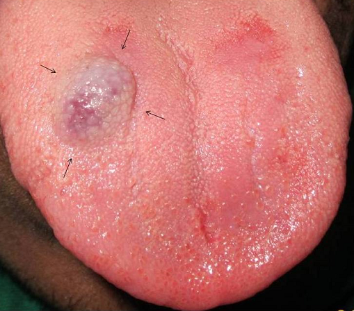
A purple swelling on the tongue
Prashanth Panta
PAMJ. 2015; 21:234. Published 31 Jul 2015 | doi:10.11604/pamj.2015.21.234.7497
Corresponding author
Prashanth Panta, Department of Oral Medicine and Radiology, MNR Dental College and Hospital, Sangareddy, Andhra Pradesh, India (maithreya.prashanth@gmail.com)
This image
| Articles published in PAMJ are Open Access and distributed under the terms of the Creative Commons Attribution 4.0 International (CC BY 4.0). |  |

eISSN: 1937-8688
The Pan African Medical Journal (ISSN: 1937-8688) is a subsidiary of the Pan African Medical Journal. The contents of this journal is intended exclusively for professionals in the medical, paramedical and public health and other health sectors.
Currently tracked by: DOAJ, AIM, Google Scholar, AJOL, EBSCO, Scopus, Embase, IC, HINARI, Global Health, PubMed Central, PubMed/Medline, ESCI
Physical address: "Kenya: 3rd Floor, Park Suite Building, Parkland Road, Nairobi. PoBox 38583-00100, tel: +254 (0)20-520-4356 | Cameroon: Immeuble TechnoPark Essos, Yaounde, PoBox: 10020 Yaounde, tel: +237 (0)24-309-5880"

For authors
Recently in the PAMJ Blog
About PAMJ - Manuscript Hut™
The Manuscript Hut is a product of the PAMJ Center for Public health Research and Information.
Kenya: 3rd Floor, Park Suite Building, Parkland Road, Nairobi. PoBox 38583-00100, tel: +254 (0)20-520-4356
Cameroon: Immeuble TechnoPark Essos, Yaounde, PoBox: 10020 Yaounde, tel: +237 (0)24-309-5880
Uganda: African Field Epidemiology network,Wings B & C, Ground Floor, Lugogo House, Plot 42, Lugogo By-pass, Kampala
Copyright © - Pan African Medical Journal - CEPHRI. 2026
Haraka Publishing Platform - (MMS V.2.5). Release date Jan 2018 - Customized for The Pan African Medical Journal
For advertisers Contact the PAMJ sales service. Download our latest media-kit.
sales-service@panafrican-med-journal.com
 |