
Case report: incidental findings of COVID-19 infection on positron emission tomography/computed tomography for staging of a giant gastric gastrointestinal stromal tumor
Hamish Reed-Embleton, Khurram Shahzad Khan, Navin Mathias, Sajid Mahmud
Corresponding author: Khurram Shahzad Khan, Department of Surgery, University Hospital Hairmyres, East Kilbride, Scotland, UK 
Received: 29 Apr 2020 - Accepted: 01 May 2020 - Published: 11 May 2020
Domain: Nuclear medicine,Radiology
Keywords: Covid-19, FDG-PET, GIST, gastrointestinal stromal tumour
This article is published as part of the supplement PAMJ Special issue on COVID - 19 in Africa, commissioned by The Pan African Medical Journal.
©Hamish Reed-Embleton et al. Pan African Medical Journal (ISSN: 1937-8688). This is an Open Access article distributed under the terms of the Creative Commons Attribution International 4.0 License (https://creativecommons.org/licenses/by/4.0/), which permits unrestricted use, distribution, and reproduction in any medium, provided the original work is properly cited.
Cite this article: Hamish Reed-Embleton et al. Case report: incidental findings of COVID-19 infection on positron emission tomography/computed tomography for staging of a giant gastric gastrointestinal stromal tumor. Pan African Medical Journal. 2020;35(2):28. [doi: 10.11604/pamj.supp.2020.35.2.23167]
Available online at: https://www.panafrican-med-journal.com//content/series/35/2/28/full
Case report 
Case report: incidental findings of COVID-19 infection on positron emission tomography/computed tomography for staging of a giant gastric gastrointestinal stromal tumor
Case report: incidental findings of COVID-19 infection on positron emission tomography/computed tomography for staging of a giant gastric gastrointestinal stromal tumor
Hamish Reed-Embleton1, Khurram Shahzad Khan1,&, Navin Mathias2, Sajid Mahmud1
1Department of Surgery, University Hospital Hairmyres, East Kilbride, Scotland, UK, 2Department of Radiology, University Hospital Hairmyres, East Kilbride, Scotland, UK
&Corresponding author
Khurram Shahzad Khan, Department of Surgery, University Hospital Hairmyres, East Kilbride, Scotland, UK
We report the incidental finding of COVID-19 in a 59-year-old male, with no significant cardiorespiratory past medical history who underwent a fluorodeoxyglucose positron emission tomography (FDG-PET) scan for investigation of a likely gastric gastrointestinal stromal tumor (GIST). There may be significant discrepancies between clinical symptoms and radiological severity with COVID-19 infection. FDG-PET scanning has the potential to complement traditional radiological imaging in COVID-19 in diagnosis of subclinical diagnosis or early stage disease, as well as monitoring disease progression.

COVID-19 was first reported in Wuhan, Hubei province, China in December 2019 [1]. In the UK, the first case of COVID-19 was reported on 31st January 2020. There are over 150,000 confirmed cases in the UK with over 20,000 reported deaths by the end of April 2020 [2]. COVID-19 symptoms can be non-specific ranging from no symptoms to severe pneumonia and death. RT-PCR remains the gold standard investigation but has high false negative rates [3]. Plain chest X-ray films may also demonstrate normal findings in the early stages of infection and are not sensitive in the detection of ground glass opacities [4]. This report emphasises the need to remain vigilant as asymptomatic carriers of COVID-19 may undergo nuclear medicine investigation.

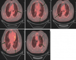
A 59-year-old male with no cardiorespiratory or other co-morbidities was referred for investigation of intermittent dysphagia. He underwent upper gastrointestinal (GI) endoscopy. This showed possible gastrointestinal stromal tumour (GIST) in the proximal stomach, although biopsies on two occasions showed normal gastric mucosa. A computed tomography (CT) scan performed in March 2020 which showed a heterogenous mass intimately connected to the gastric fundus, suggestive of gastric GIST and measuring 15 x 12.6cm. Discussion within a regional sarcoma multidisciplinary team suggested referral for endoscopic ultrasound (EUS) and functional imaging with fluorodeoxyglucose positron emission tomography (FDG-PET). The FDG-PET scan was performed four weeks later. At this stage he remained apyrexial (body temperature < 37.50C), with no respiratory or gastrointestinal symptoms, or any headache and there was no history of exposure to possible COVID-19. The FDG-PET scan confirmed the CT findings and reported no significant change or size in the GIST but showed FDG-avid, prominently peripheral, ground-glass changes in bilateral upper and lower lobes (Figure 1). The changes were new compared with the CT scan dated 20th March 2020 and were in keeping with probable COVID-19 infection [1,5,6]. His FDG-PET scan results were discussed via phone call and he was advised to self-isolate. He remains asymptomatic.
Our case again highlights the speed of disease progression and dramatic radiological findings despite the apparent lack of symptoms in keeping with recent data that suggests up to 78% of patients with a COVID-19 infection may be asymptomatic [7,8]. Viral infection causes an inflammatory cascade during which all activated leucocytes are dependent on glucose for anaerobic glycolysis which causes a high FDG uptake during FDG-PET scanning [3,9]. Qin et al., reported FDG-PET scans of four confirmed or suspected COVID-19 patients [10]. All lesions showed high trace uptake (SUVmax 1.8-12.2), demonstrating the high inflammatory burden associated with COVID-19 infection and noted that patients with higher FDG uptake took longer to heal [10]. This supports the potential use of FDG-PET scanning to aid in determining the progression, and outcomes in patients with COVID-19.
Chefer et al. reported the ability of FDG-PET to detect even subtle changes with subclinical MERS-CoV infection in 2015 [11]. Such findings indicate the potential added value of FDG-PET imaging when presentation, is non-specific or atypical. Interestingly FDG-PET scanning is an important tool for investigating fever of unknown origin and measuring the inflammatory response to infection [9]. Radiologic imaging techniques such as CT scan are able to detect focal inflammatory and infectious processes. However, during the early phase of infection, the anatomical change may be insubstantial for their detection by CT scan compared with FDG-PET scan [9]. This is supported by Bellani et al., who reported, in patients with acute respiratory distress syndrome (ARDS), FDG-PET imaging has been shown to identify the increase metabolic rates of pulmonary tissues when CT scanning finds them to be normal [12]. FDG-PET scanning has further been shown to aid in the prediction of patients developing ARDS following thoracic trauma and pulmonary contusion [13].
With a relatively low radiation exposure, radionucleotide imaging may be able to give a more complete picture of acute respiratory viral infections and the development of pulmonary parenchymal injury and may enable more logical decision making around use of specific interventions such as the use of anti-inflammatories [14,15]. Therefore, it has the potential to complement CT scan and other imaging modalities in identifying the pulmonary structural changes, evaluating infectious and immune phases and monitoring disease progression during the course of acute respiratory illness with COVID-19 infection [14,16]. However, in the current era of financial strain this test is not feasible or readily available for diagnosing COVID-19.
The authors declare no competing interests.
Hamish Reed-Embleton: conception and design, acquisition, analysis & interpretation of data, drafting of work and final approval; Khurram Shahzad Khan: conception and design, acquisition, analysis & interpretation of data, drafting of work and final approval; Navin Mathias: acquisition of data, drafting of work and final approval; Sajid Mahmud: conception and design, drafting of work and final approval. All the authors have read and agreed to the final manuscript.
Figure 1: CT PET showing faintly FDG-avid patchy prominently peripheral ground-glass changes in bilateral upper and lower lobes in keeping with probable COVID-19 infection
- Zu ZY, Jiang M Di, Xu PP, Chen W, Ni QQ, Lu GM et al. Coronavirus disease 2019 (COVID-19): a perspective from China. Radiology. 2020;200490. PubMed | Google Scholar
- Johns Hopkins Coronavirus Resource Center. New Cases of COVID-19 In World Countries. 2020. Accessed Apr 24 2020
- Deng Y, Lei L, Chen Y, Zhang W. The potential added value of FDG PET/CT for COVID-19 pneumonia. Eur J Nucl Med Mol Imaging. 2020;19-20. PubMed | Google Scholar
- Ming-Yen Ng, Elaine YPL, Jin Y, Fangfang Y, Xia L, Hongxia W et al. Imaging Profile of the COVID-19 Infection: radiologic findings and literature review. Radiologic. 2020;2(1). Google Scholar
- Zhou S, Wang Y, Zhu T, Xia L. CT features of coronavirus disease 2019 (COVID-19) pneumonia in 62 patients in Wuhan, China. Am J Roentgenol. 2020:1-8. PubMed | Google Scholar
- Kanne JP, Little BP, Chung JH, Elicker BM, Ketai LH. Essentials for radiologists on COVID-19: an update-radiology scientific expert panel. Radiology. 2020;200527. PubMed | Google Scholar
- WHO. Report of the WHO-China joint mission on coronavirus disease 2019 (COVID-19). WHO-China Jt Mission Coronavirus Dis 2019. 2020;2019(February):16-24.
- Day M. Covid-19: four fifths of cases are asymptomatic, China figures indicate. BMJ. 2020;369:m1375. PubMed | Google Scholar
- Kouijzer IJE, Mulders-Manders CM, Bleeker-Rovers CP, Oyen WJG. Fever of unknown origin: the value of FDG-PET/CT. Semin Nucl Med. 2018;48(2):100-7. PubMed | Google Scholar
- Qin C, Liu F, Yen TC, Lan X. 18F-FDG PET/CT findings of COVID-19: a series of four highly suspected cases. Eur J Nucl Med Mol Imaging. 2020;47(5):1281-6. PubMed | Google Scholar
- Chefer S, Thomasson D, Seidel J, Reba RC, Bohannon JK, Lackemeyer MG et al. Modeling [18F]-FDG lymphoid tissue kinetics to characterize nonhuman primate immune response to Middle East respiratory syndrome-coronavirus aerosol challenge. EJNMMI Res. 2015 Dec;5(1):65. PubMed | Google Scholar
- Bellani G, Messa C, Guerra L, Spagnolli E, Foti G, Patroniti N et al. Lungs of patients with acute respiratory distress syndrome show diffuse inflammation in normally aerated regions: a [18F]-fluoro-2-deoxy-D-glucose PET/CT study. Crit Care Med. 2009;37(7):2216-22. PubMed | Google Scholar
- Rodrigues RS, Miller PR, Bozza FA, Marchiori E, Zimmerman GA, Hoffman JM et al. FDG-PET in patients at risk for acute respiratory distress syndrome: a preliminary report. Intensive Care Med. 2008;34(12):2273-8. PubMed | Google Scholar
- Kash JC. Applications of high-throughput genomics to antiviral research: evasion of antiviral responses and activation of inflammation during fulminant RNA virus infection. Antiviral Research. 2009. PubMed | Google Scholar
- Bray M, Lawler J, Paragas J, Jahrling PB, Mollura DJ. Molecular imaging of influenza and other emerging respiratory viral infections. J Infect Dis. 2011;203(10):1348-59. PubMed | Google Scholar
- Guedj E, Verger A, Cammilleri S. PET imaging of COVID-19: the target and the number. Eur J Nucl Med Mol Imaging. 2020 Apr 17. PubMed | Google Scholar
Search
This article authors
On Pubmed
On Google Scholar
Citation [Download]
Navigate this article
Similar articles in
Key words
Tables and figures
This supplement
Article metrics
Recently from the JOURNAL_ABBREVIATION


Authors´ services
