
Delayed hypersensitivity as a pathophysiological mechanism in cutaneous lesions due to SARS-CoV-2
Mostafa Rafai, Jalal Elbenaye, Sana Sabry, Hicham Janah
Corresponding author: Mostafa Rafai, Department of Physiology, Faculty of Medicine and Pharmacy, Hassan II University, Casablanca, Morocco 
Received: 13 Jul 2020 - Accepted: 15 Jul 2020 - Published: 16 Jul 2020
Domain: Dermatology
Keywords: COVID-19, chilblains, purpura, erythema multiforme, hypersensitivity
This article is published as part of the supplement PAMJ Special issue on COVID - 19 in Africa, commissioned by The Pan African Medical Journal.
©Mostafa Rafai et al. Pan African Medical Journal (ISSN: 1937-8688). This is an Open Access article distributed under the terms of the Creative Commons Attribution International 4.0 License (https://creativecommons.org/licenses/by/4.0/), which permits unrestricted use, distribution, and reproduction in any medium, provided the original work is properly cited.
Cite this article: Mostafa Rafai et al. Delayed hypersensitivity as a pathophysiological mechanism in cutaneous lesions due to SARS-CoV-2. Pan African Medical Journal. 2020;35(2):115. [doi: 10.11604/pamj.supp.2020.35.2.24980]
Available online at: https://www.panafrican-med-journal.com//content/series/35/2/115/full
Letter to the editors 
Delayed hypersensitivity as a pathophysiological mechanism in cutaneous lesions due to SARS-CoV-2
Delayed hypersensitivity as a pathophysiological mechanism in cutaneous lesions due to SARS-CoV-2
Mostafa Rafai1,&, Jalal Elbenaye2,3, Sana Sabry1, Hicham Janah4
&Corresponding author

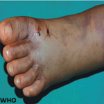
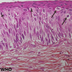
A 17-year-old adolescent with no medical history; documented to have a mild SARS-CoV-2 infection (clinical symptoms and minimal peripheral ground-glass opacities in both lungs in chest CT); had chilblains-like lesions on the toes (Figure 1 A) and asymptomatic erythematopurpuric lesions of soles (Figure 1 B) on the fourth day of the onset of COVID-19 symptoms. He took vitamin C only. There were no thrombocytopenia, no hypercoagulability except a slight increase of inflammatory markers. Sars-cov-2 RT-PCR was negative. On the fifteenth day of the onset of symptoms, he developed mild itching and painless erythematous maculopapular lesions of heels (Figure 1 C) with targetoid aspect on the palms (Figure 1 D). There was no mucosal involvement. No recent episode of recurrent herpes or drugs intake were noted. Reported COVID-19 associated cutaneous manifestations are various. Some occur early as exanthem, urticaria, chickenpox like rash; mainly affecting the trunk [1]; while others appear later like chilblains and maculopapular lesions with acral distribution [2]. This suppose that there would be two types of lesions according to two different pathophysiological mechanisms: first and early one which would be linked to viremia and a second; late; related to immunological and inflammatory response during the disease.
Our patient had presented chilblains-like lesions and acral purpura concomitantly, followed few days later by maculopapular lesions with targetoid lesions reminiscent of erythema multiforme. Same presentations were reported: 02 cases with chilblains-like lesions evolving to erythemato-papular targetoid lesions [3]; maculopapular lesions in heels [4]. All these observations were seen in healthy young patients, with negative SARS-CoV-2 RT-PCR, appear late and would have a good prognosis. These findings suggest that acral lesions would be the clinical expression of type III and/or IV hypersensitivity targeting the small vessels of skin then responsible for endothelial activation, dermal and perivascular lymphoid infiltrate. Histological observations corroborate this hypothesis [2-6]. These suggestions require more investigation by means of SARS-CoV-2 serological tests, more relevant histology with immunohistochemistry and immunofluorescence and finally a serum assay of complement and immunological factors.
The authors declare no competing interests.

Mostafa Rafai, Jalal Elbenaye, Sana Sabry and Hicham Janah :study, conception and design, drafting of the manuscript and critical revision. All authors have read and agreed to the final version of this manuscript.
Figure 1: A) chilblains-like lesions on the toes; B) erythematopurpuric lesions of soles; C) erythematous maculopapular lesions of the heel; D) targetoid aspect on the palms
- Recalcati S. Cutaneous manifestations in COVID-19: a first perspective. J Eur Acad Dermatol Venereol. 2020 May;34(5):e212-e213. PubMed | Google Scholar
- Kolivras A, Dehavay F, Delplace D, Feoli F, Meiers I, Milone L et al. Coronavirus (COVID-19) infection-induced chilblains: a case report with histopathological findings. JAAD Case Rep. 2020 Apr 18;6(6):489-492. PubMed | Google Scholar
- Recalcati S, Barbagallo T, Frasin LA, Prestinari F, Cogliardi A, Provero MC et al. Acral cutaneous lesions in the Time of COVID-19. J Eur Acad Dermatol Venereol. 2020 Apr 24;10.1111/jdv.16533. PubMed | Google Scholar
- Estébanez A, Pérez-Santiago L, Silva E, Guillen-Climent S, García-Vázquez A, Ramón MD. Cutaneous manifestations in COVID-19: a new contribution. J Eur Acad Dermatol Venereol. 2020 Jun;34(6):e250-e251. PubMed | Google Scholar
- Magro C, Mulvey JJ, Berlin D, Nuovo G, Salvatore S, Harp J et al. Complement associated microvascular injury and thrombosis in the pathogenesis of severe COVID-19 infection: a report of five cases. Transl Res. 2020 Jun;220:1-13. PubMed | Google Scholar
- Roncati L, Ligabue G, Fabbiani L, Malagolia C, Gallo G, Lusenti B et al. Type 3 hypersensitivity in COVID-19 vasculitis. Clinical Immunology. 2020 Aug;217:108487. PubMed | Google Scholar
Search
This article authors
On Pubmed
On Google Scholar
Citation [Download]
Navigate this article
Similar articles in
Key words

This supplement
Article metrics
Recently from the PAMJ
Authors´ services
