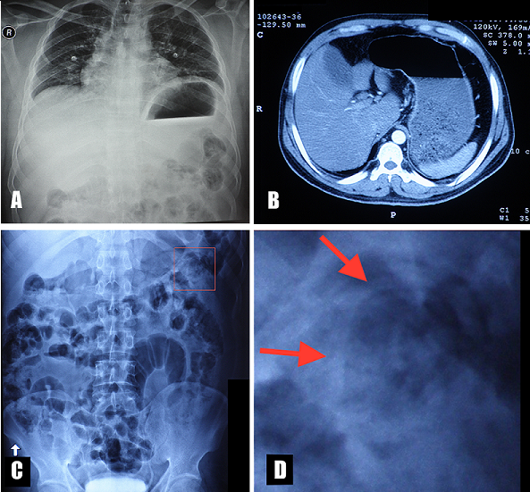
Figure 1: A) chest X-ray showing stomach dilation, prominent gastric air bubble and gastric air-fluid level; B) abdominal CT showing stomach dilation; C) erect abdominal plain X-ray with visible worms in the left upper quadrant (red square); D) detail of panel C (red square) showing tubular or cord-like curvilinear soft tissue densities in a jejunal loop characteristic of ascaris worms (red arrows)